
Learning Objectives
7 objectivesBy the end of this note, you should be able to:
- Understand the importance of water as a solvent in transport, including its dipole nature.
- Distinguish between monosaccharides, disaccharides and polysaccharides, including glycogen, amylose and amylopectin.
- Relate carbohydrate structures to their roles in providing and storing energy.
- Use a semi-quantitative method with Benedict’s reagent and iodine solution to estimate sugar and starch concentrations.
- Describe the formation of glycosidic bonds via condensation reactions and their breakdown via hydrolysis.
- Describe how a triglyceride is synthesised through condensation reactions forming ester bonds.
- Distinguish between saturated and unsaturated lipids.
Water as a Solvent in Transport
Water acts as a vital biological solvent because its dipole nature allows it to dissolve charged or polar substances and transport them around organisms.
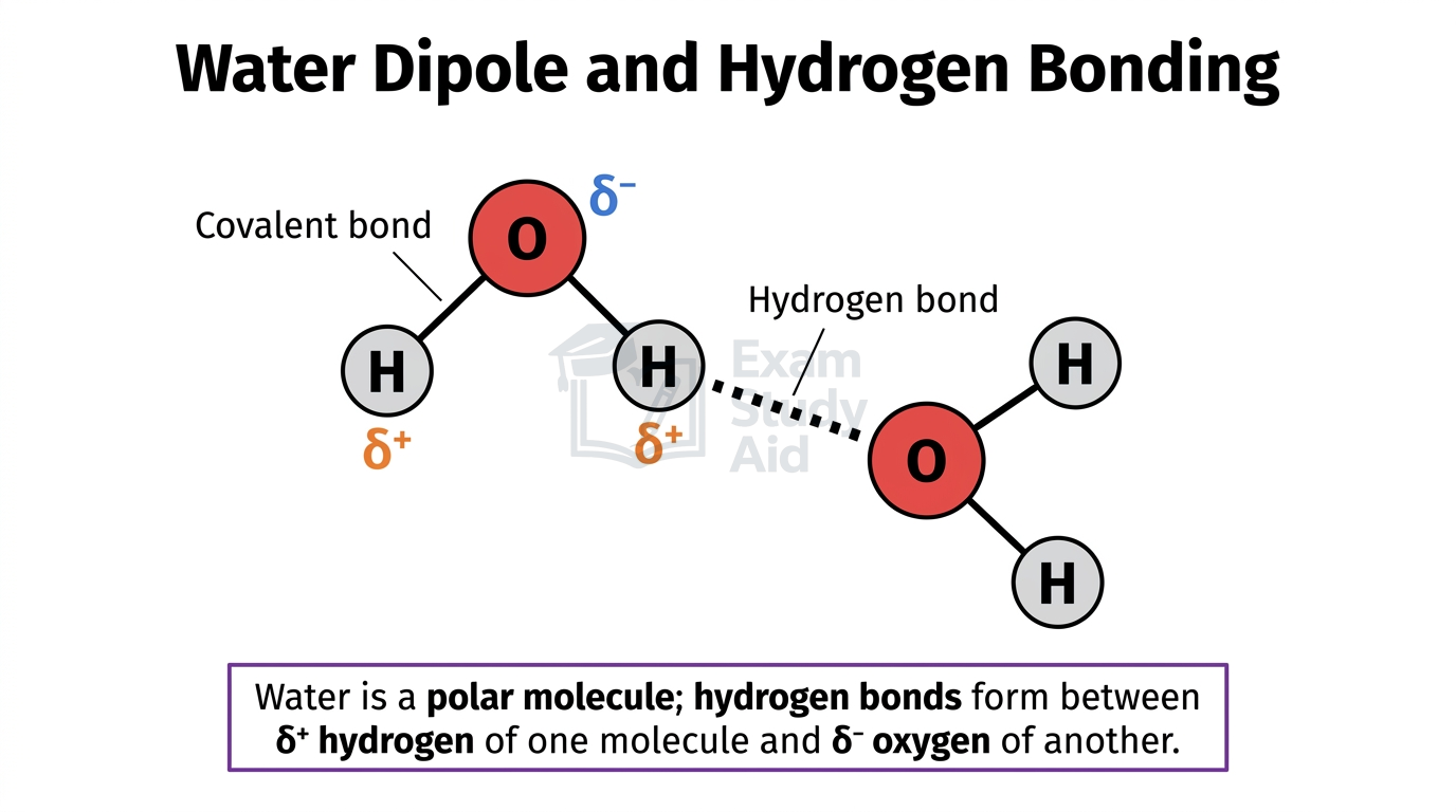
The unequal sharing of electrons between oxygen and hydrogen makes water a polar molecule. Oxygen pulls the shared electrons more strongly, becoming slightly negative (δ⁻). Each hydrogen becomes slightly positive (δ⁺). This permanent separation of charge across the molecule is called a dipole.
When ionic or polar solutes enter water, the dipoles attract them. The slightly positive hydrogens face negative ions. The slightly negative oxygens face positive ions. Water molecules surround each solute particle and pull them apart, dissolving them.
This solvent property is essential for transport in living organisms. Glucose, amino acids, mineral ions, hormones, and urea all dissolve in blood plasma. The circulatory system can therefore carry them around the body. In plants, mineral ions and sucrose dissolve in water for transport through xylem and phloem. Inside cells, the watery cytoplasm provides the medium for metabolic reactions to occur.
Hydrogen bonds also form between adjacent water molecules. These weak attractions occur between the δ⁺ hydrogen of one molecule and the δ⁻ oxygen of another. The resulting cohesion supports the continuous water column drawn up xylem during transpiration.
In contrast, non-polar substances like lipids are hydrophobic and do not dissolve in water. They are transported by proteins, for example as lipoproteins in blood.
Classifying Carbohydrates and Energy Roles
Carbohydrates are organic molecules built from carbon, hydrogen, and oxygen, classified by size into three groups: monosaccharides, disaccharides, and polysaccharides.
Monosaccharides are single sugar units, typically with the general formula (CH₂O)ₙ. The three required examples are glucose, fructose, and galactose. They are small, polar, and soluble, which makes them easy to transport in solution. Glucose is the main respiratory substrate: it is broken down to release energy in cells.
Disaccharides form when two monosaccharides join through condensation. The three required examples are maltose (glucose + glucose), sucrose (glucose + fructose), and lactose (glucose + galactose). They remain soluble and act as transport sugars. For example, sucrose is the main transport sugar in plant phloem.
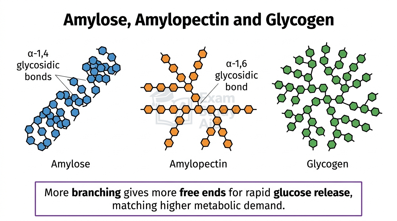
Polysaccharides are long chains or branched structures made of many monosaccharide units. They are insoluble, so they do not affect water potential or osmotic balance inside cells. This insolubility, combined with their compact shape, makes them ideal energy storage molecules. The three required examples are glycogen, amylose, and amylopectin.
| Polysaccharide | Found in | Structure | Bonds present | Role |
|---|---|---|---|---|
| Amylose | Plants (in starch) | Linear, coiled into a helix | α-1,4 glycosidic | Compact glucose storage |
| Amylopectin | Plants (in starch) | Branched | α-1,4 and α-1,6 glycosidic | Faster glucose release through many branch ends |
| Glycogen | Animals (liver and muscle) | Highly branched, more than amylopectin | α-1,4 and α-1,6 glycosidic | Very rapid glucose release for high metabolic demand |
The link between structure and function is consistent. The helix in amylose makes it compact for storage. Branching gives many ends for enzymes to act on simultaneously, allowing fast glucose release. Glycogen is the most branched because animals have higher metabolic rates than plants and need glucose mobilised quickly. Insolubility means these large molecules do not lower the water potential of storage cells, preventing unwanted osmotic water uptake.
Testing for Reducing Sugars and Starch
Two important biochemical tests allow semi-quantitative estimation of carbohydrate concentrations: Benedict’s reagent detects reducing sugars, while iodine solution detects starch.
Benedict’s reagent contains copper(II) sulfate. When heated with a reducing sugar, the sugar donates electrons that reduce Cu²⁺ to Cu⁺. This produces an insoluble brick-red precipitate of copper(I) oxide. As the concentration of reducing sugar increases, more precipitate forms and the colour shifts from blue → green → yellow → orange → brick-red.
Reducing sugars include all monosaccharides (glucose, fructose, galactose) and most disaccharides (maltose, lactose). Sucrose is non-reducing and produces no colour change directly with Benedict’s. To test for sucrose, the sample must first be hydrolysed (e.g. with dilute hydrochloric acid) to release reducing monosaccharides.
Iodine solution (potassium iodide with iodine) detects starch. When iodine enters the helical coils of amylose, the starch–iodine complex absorbs light strongly, producing a blue-black colour. The deeper the colour, the more starch present.
Both tests can be used semi-quantitatively by comparing samples to a series of solutions of known concentration (colour standards).
MisconceptionMany students assume sucrose gives a positive Benedict’s test simply because it is a sugar. Sucrose is non-reducing and gives no colour change unless first hydrolysed. Maltose and lactose, however, are reducing and do react.
Exam TipAlways check whether the named sugar is reducing before predicting a Benedict’s result.
CORE PRACTICAL 1: Estimating Sugar and Starch Concentrations Using Colour Standards
Aim: To estimate the concentration of reducing sugars and starch in unknown samples by comparing colours to a set of standards.
- Prepare a series of glucose solutions of known concentration, for example 0%, 0.5%, 1.0%, 2.0%, and 5.0%.
- Add an equal volume of Benedict’s reagent to each tube.
- Place all tubes in an 80°C water bath for the same time, for example five minutes.
- Record (or photograph) the colour of each sample after heating to produce a set of colour standards.
- Test an unknown sample under identical conditions and compare its colour to the standards to estimate concentration.
- To test for starch, add iodine solution to a series of starch standards of known concentration, then compare an unknown sample.
- Independent variable (IV): concentration of reducing sugar or starch in the standard solutions.
- Dependent variable (DV): colour produced after the test, judged visually or measured with a colorimeter.
- Control variables (CV): volume of reagent, sample volume, water-bath temperature, heating time, lighting when comparing colours.
Benedict’s progresses from blue (no reducing sugar) through green, yellow, and orange to brick-red (high concentration). Iodine progresses from orange-brown (no starch) to blue-black (high starch). Matching the unknown’s colour to a standard estimates its concentration.
Visual colour matching is subjective and varies between observers. Use a colorimeter to measure absorbance at a fixed wavelength and compare to a calibration curve for an objective, accurate result. Also ensure all tubes have identical heating temperature and time, so colour reflects only concentration.
Glycosidic Bonds, Condensation and Hydrolysis
Monosaccharides join through condensation reactions to form disaccharides and polysaccharides, creating glycosidic bonds that are later broken by hydrolysis when sugars are needed.
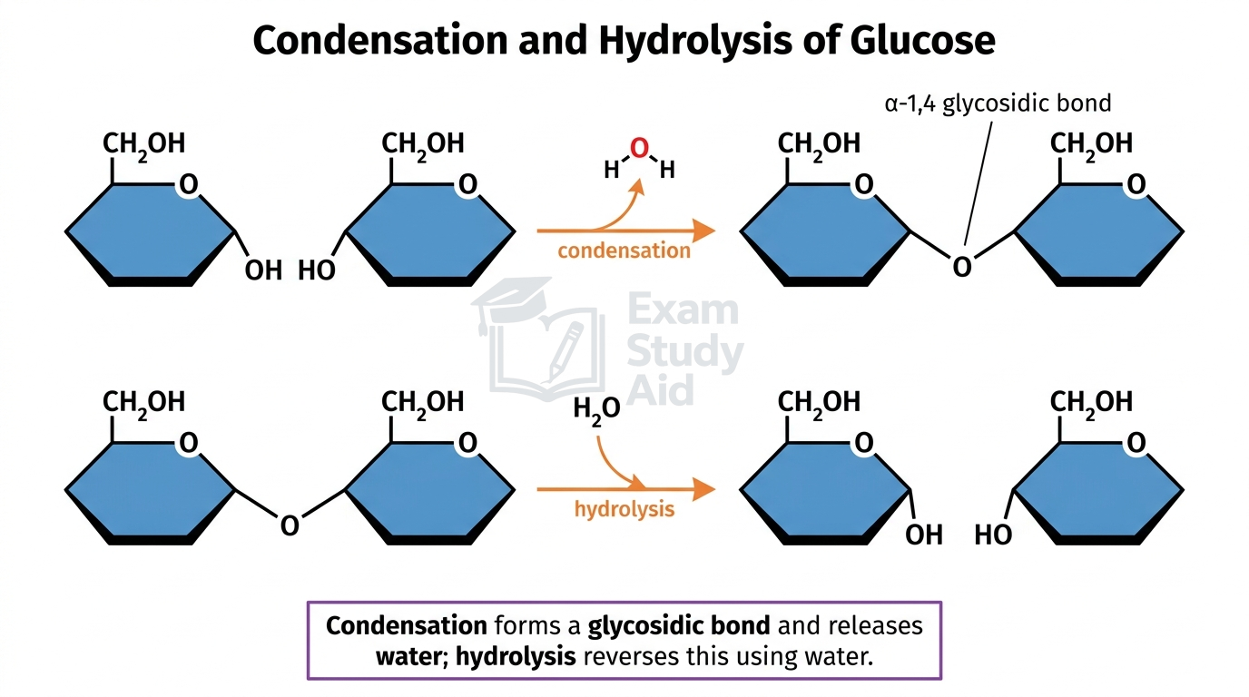
In a condensation reaction, two monosaccharides come together. A hydroxyl (–OH) group on one sugar reacts with a hydroxyl group on the next. A molecule of water (H₂O) is released, and a new covalent glycosidic bond links the two sugars. For example, an α-1,4 glycosidic bond joins carbon 1 of one glucose to carbon 4 of the next.
Examples of disaccharide formation:
- glucose + glucose → maltose + H₂O
- glucose + fructose → sucrose + H₂O
- glucose + galactose → lactose + H₂O
Polysaccharides such as glycogen, amylose, and amylopectin form by repeated condensation reactions. Each step releases one water molecule and forms one glycosidic bond.
In a hydrolysis reaction, a water molecule is added across a glycosidic bond, breaking it. The –H from water joins one sugar, and the –OH joins the other, releasing the original monosaccharides. Hydrolysis is catalysed by specific enzymes. Amylase hydrolyses starch into maltose, and maltase hydrolyses maltose into glucose. Hydrolysis is essential during digestion and during the breakdown of stored polysaccharides for respiration.
Examiner Insight“Glycosidic bond” earns the mark; “chemical bond” or “sugar bond” does not. For full credit, name the specific bond (glycosidic) and the type of reaction (condensation or hydrolysis), and state that water is released or added.
Exam TipAlways pair the bond name with the reaction type and the role of water.
Triglycerides and Lipid Saturation
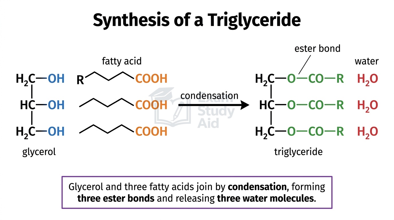
A triglyceride is a major lipid formed when one glycerol combines with three fatty acids through condensation reactions, producing three ester bonds.
Each fatty acid joins glycerol when its carboxyl group (–COOH) reacts with a hydroxyl group (–OH) on the glycerol. One water molecule is released per bond formed, so each triglyceride synthesis releases three water molecules in total. The bond formed is called an ester bond, characterised by the C(=O)–O linkage between the fatty acid and glycerol.
Triglycerides are excellent energy stores. They contain many C–H bonds, releasing approximately twice the energy per gram as carbohydrate when respired. They are insoluble, so they do not affect water potential. Stored compactly in adipose tissue, they also provide thermal insulation, cushioning of organs, and (when respired) metabolic water.
Triglycerides can be saturated or unsaturated, depending on the bonds in their fatty acid chains:
| Property | Saturated | Unsaturated |
|---|---|---|
| Bonds in hydrocarbon chain | All single C–C bonds | At least one C=C double bond |
| Shape of chain | Straight | Kinked at each double bond |
| Packing | Pack tightly together | Pack loosely due to kinks |
| State at room temperature | Solid (e.g. butter, lard) | Liquid (e.g. olive oil, sunflower oil) |
| Typical source | Animal fats | Plant oils, fish oils |
A fatty acid with one C=C double bond is monounsaturated, while one with multiple C=C bonds is polyunsaturated.
QUICK RECAP
Key Points
- Water is polar — oxygen δ⁻, hydrogens δ⁺.
- Hydrogen bonds form between adjacent water molecules.
- Polar and ionic solutes dissolve due to attraction with water dipoles.
- Water transports glucose, ions, hormones, and urea in blood plasma.
- Monosaccharides include glucose, fructose, and galactose.
- Disaccharides: maltose (G+G), sucrose (G+F), lactose (G+Gal).
- Polysaccharides: glycogen, amylose, amylopectin — insoluble and compact.
- Amylose is unbranched and helical, contains α-1,4 glycosidic bonds.
- Amylopectin is branched, with α-1,4 and α-1,6 glycosidic bonds.
- Glycogen is highly branched; found in animal liver and muscle.
- Branching gives many ends for fast glucose mobilisation.
- Glycosidic bonds form during condensation, releasing one water molecule.
- Hydrolysis adds water across bonds, breaking them into monomers.
- Benedict’s: blue → brick-red with reducing sugars when heated.
- Iodine: orange-brown → blue-black with starch.
- Sucrose is non-reducing — no Benedict’s colour change directly.
- Triglyceride = glycerol + 3 fatty acids, joined by 3 ester bonds.
- Saturated lipids: only single C–C bonds, solid at room temperature.
- Unsaturated lipids: contain C=C double bonds, kinked, liquid at room temperature.
CAN I…? PROGRESS CHECK
Self-Assessment
- Explain why water acts as a solvent for ionic and polar substances?
- Give examples of substances transported in solution in animals and plants?
- Distinguish monosaccharides, disaccharides, and polysaccharides with examples?
- Relate the structures of glycogen, amylose, and amylopectin to their roles in energy storage?
- Use Benedict’s reagent and colour standards to estimate reducing sugar concentration?
- Use iodine solution and colour standards to estimate starch concentration?
- Explain why sucrose gives no colour change with Benedict’s reagent directly?
- Describe the formation of glycosidic bonds via condensation and breakdown via hydrolysis?
- Describe how a triglyceride is synthesised from glycerol and three fatty acids?
- Distinguish between saturated and unsaturated lipids by structure and properties?